Clemson students study swine in search of answers to breast cancer
CLEMSON — A passion born of a personal connection to the ravages of breast cancer is pushing a team of Clemson University Animal and Veterinary Sciences students to participate in an innovative undergraduate research project that could lead to discovery, advance their careers and propel them to the world stage.

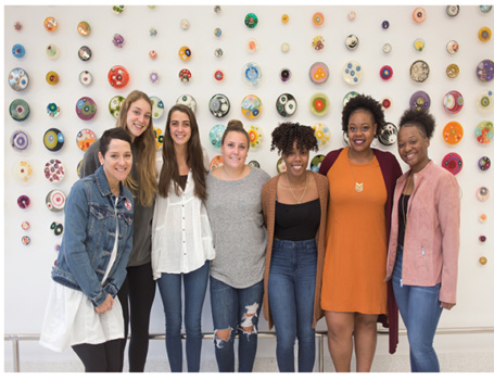
The group consists of Sabrina Carrel of Poway, California; Paula Lewis of Marion; Hannah Oswalt of Long Island; Brooke Redmond of Columbia; Shelby Smith of Huntsville, Alabama and Amber Stone of Columbia, and is led by Heather Dunn, a senior lecturer in the Clemson Animal and Veterinary Sciences Department.
They are participating in the Bioinformatics for Cancer Genomics Creative Inquiry project. Their research uses a novel approach to study the mechanism that causes mammary glands to develop as a way to understand and possibly halt the growth of aggressive breast cancer cells.
“From the time we are conceived, through birth and up to puberty, our cells are changing, growing and establishing new patterns of signals,” Dunn said. “This cell conversion is called the epithelial-mesenchymal transition, or EMT. When this transition occurs, cells have the ability to migrate to other parts of the body and divide. Research confirms that this process also occurs during cancer and is believed to be the early initiating step in the development of triple negative breast cancer, or TNBC.”
The students are using a minimally invasive biopsy technique invented by Dunn and patented by Clemson University, as well as an array of Clemson University laboratories and imaging facilities to study the mammary cells of prepubescent swine to unlock the mystery of EMT in cancer. The ultimate goal is to someday be able to cease the EMT process in triple negative breast cancer cells.
The swine genes are compared to genes found in human breast cancer profiles. Previous studies used mice samples, but swine mammary gland tissue more closely resembles human mammary gland tissue. Tissue samples are collected using the Dunn Biopsy Method and analyzed in various Clemson laboratories.
This research has been underway for only two semesters, but cancer research experts already are taking note of the students’ study. The students have been invited to present at the Keystone Symposia of Molecular and Cellular Biology conference in Florence, Italy, March 15-19.
Utilizing Clemson resources
Students in this project are introduced to Clemson University resources few undergraduates know exists including: The Histology Laboratory, the Clemson Light Imaging Facility (CLIF), as well as programs for bioinformatic analyses.
Field and lab work begin when mammary gland genes are collected from pigs at Clemson’s Starkey Swine Center using the Dunn Biopsy Method. This method was created by Dunn and currently filed as a provisional patent through the Clemson University Research Foundation.
Once samples are collected, multiple laboratories are used to process the tissue. The Animal and Veterinary Science Histology Core Facility is where the students prepare the samples for analysis.
“This lab is where we embed the tissue samples in paraffin wax to preserve the tissue for storage and later use,” Redmond said. “It is also where we slice the samples and put them on slides just before we stain them so that they can be observed under a microscope. We have to be able to analyze samples we’ve collected and what we’re doing in this lab allows us to do that.”

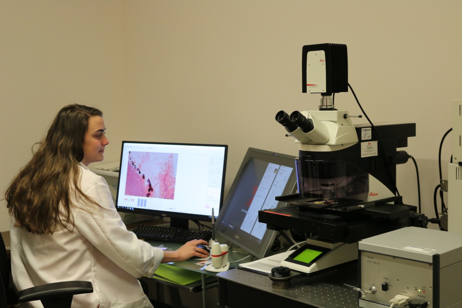
Shelby Smith of Huntsville, Alabama, uses equipment in Clemson’s Light Imaging Facility to view slides from her mother’s breast biopsy and compare human cells to swine cells.
In the Clemson Light Imaging Facility, cells are selected and extracted using a laser microdissection microscope for analyses.
“We use the microscope to laser cut out different cell populations so that we can determine molecular events from different populations of cells,” Smith said. “This will help us understand how the cells communicate with their environment.”
Smith’s mother died from breast cancer in 2003. She found slides from her mother’s biopsies and is including these slides in the Creative Inquiry study. Equipment in the Light Imaging Facility is used to view the slides and compare human cells to swine cells.
“It’s helpful to be able to see real human cancer cells,” Smith said. “My mom died when I was five and ever since then I just really want to know about it and I want to cure cancer.”
Once cell samples have been collected and data compiled, the students use bioinformatics to analyze their data. Programs such as the National Cancer Institute’s Genomic Data Commons Data Portal (GDC) are used to compare the swine mammary genes to human mammary genes. This portal allows interactive analysis and data visualization specifically identifying gene mutations, frequencies and expression networks.
Being able to use these resources as undergraduates is something Oswalt believes she will benefit from in the future. She and the other students plan to pursue careers in medicine.
“We are being exposed to multiple lab techniques such as laser dissection and biopsy sampling that we usually don’t get to see or learn in our classes,” Oswalt said. “This CI project is exposing us to bioinformatics, and I believe this will be extremely beneficial in our future as this has become a prominent field in the world of medicine and research.”
It was the opportunity to learn new things that prompted the students to enroll in the project. It’s also the project that encouraged them to continue their route in Animal and Veterinary Sciences.
“I had a new passion for human medicine,” Lewis said. “Dr. Dunn talked with me about this project and I thought this would be a great way to incorporate my current degree with my new interest.”
Stone had a similar desire.
“I became involved in this CI project after an advising appointment with Dr. Dunn,” Stone said. “I expressed to her my interest in going into the human medical field. She mentioned to me that she was going to have a CI project the following two semesters and asked if I was interested in being a part of it. She briefly explained what her CI project would be and she totally had me sold!”
Persistence pays off
Researchers across the globe are taking note of the students’ work. The entire group has been invited to present during the Keystone Symposia’s Cancer Metastasis conference in Florence, Italy, in March. The students’ presentation is titled – Evaluation of swine mammary glands: A model for development, cancer and environmental cues. The students are excited about the group being recognized for its work in cancer research.
“This is a huge and amazing opportunity,” Lewis said. “I am extremely happy that I am able to be on this team. Being invited to present at this conference lets me know that our hard work this semester will be seen by others and we can expand on our research to help work towards a cure for breast cancer.”
In addition to the vast knowledge and experiences they have gained by participating in this program, all of the students said the interest Dunn has shown in them and her willingness to help them succeed has been overwhelming.
“This CI has given me more than I can easily explain in words,” Carrel said. “I have always had outrageous dreams. I’m the college kid who wanted to study language, arts, science and medicine all at once. I’m the one people laughed at for trying very hard to accomplish that.
“Dr. Dunn was the first professor to take me seriously and to help me work towards making at least some of my dreams possible. With the tools and experience that this CI has given me I seriously feel that I can go on after Clemson to do absolutely anything I put my heart into.”
Triple negative breast cancer is the only type of breast cancer that lacks a specific Federal Drug Administration approved drug for treatment. This aggressive form of cancer has a high mortality rate and is associated with racial disparities – occurring in 23 percent of African American women and just 12 percent in other races.
To help raise awareness, education and support of triple negative breast cancer, Triple Negative Breast Cancer Day is recognized annually around the world on March 3.

